ACHILLES TENDON PAIN
There are two main conditions that affect the Achilles tendon. One is known as Achilles tendonitis (A.T) and the other is Achilles tendinosis. A.T is a painful condition that causes pain around the back of the ankle and heel area. It usually involves inflammation around the Achilles tendon or within the sheath that surrounds it.
Redness and swelling are not uncommon and the physiological change in the Achilles tendon and surrounding tissue can be seen with the naked eye. The redness and heat are associated with increased blood flow to the area or hyperaemia and this can be observed in ultrasound images. Tendon rupture is the other possible Achilles tendon injury.
TORN ACHILLES TENDON OR AN ACHILLES TENDON RUPTURE
A ruptured tendon can occur if your Achilles tendon is overstretched. This overstretching causes a tendon rupture or snap. The tendon can rupture completely or can tear partially. It is very painful and is likely to affect walking. Most of the time, non-surgical treatment can be effective in healing the Achilles tendon but it isn’t uncommon for surgery to be performed if the Achilles tendon rupture is significant.
INSERTIONAL ACHILLES TENDONITIS
Insertional A.T is the degeneration or pain near where the fibres of the Achilles tendon attach to the heel bone. This is generally treated the same way as other forms Achilles tendon injuries, such as A.T or tendinosis. Sometimes, bone spurs are seen in conjunction with insertional A.T. However it is not the bone spurs themselves that cause the heel pain, but rather the inflammation of the tendon in the area of the spur. Treating a heel spur can be a part of addressing the overall issue.
ACHILLES TENDINOSIS VS ACHILLES TENDONITIS
Achilles tendinosis is different from A.T in that there is usually an absence of inflammation. This is because tendinosis is more of a degenerative change in the Achilles tendon, including disorganisation of the tendon fibres and/or micro-tearing of the tendon. Achilles tendinosis usually occurs following a chronic bout of A.T and while the symptoms are similar there are some differences.
The treatment of these two Achilles tendon injuries should also differ and must involve careful therapeutic consideration. The symptomatic area is usually the lower section of the Achilles tendon where it attaches to the back of the heel.
ACHILLES TENDINOSIS / TENDINOSIS SYMPTOMS
Patients with a sore Achilles tendon due to A.T often feel stiffness or a tight feeling when they begin to walk (in the mornings or at other times) which “warms up” or feels better the more they move. Runners who have been diagnosed with A.T are often still able to complete their activities without too much discomfort.
This is possibly due to the increase in blood flow that follows the activity. The blood flow is thought to flush away some of the inflammatory cells which “frees up” the tendon and reduces the heel pain.
The symptoms return not long after the run and can be a problem the following morning when the patient rises from bed and begins to walk. Once again, the increase in blood flow which follows movement and possibly a warm shower reduces the symptoms and the patient is able to walk more freely again.
Patients with an Achilles tendon injury will feel a sharp pain when pressure is applied to the sides of the tendon with a pinching effect, for example with finger and thumb. The painful area can reside anywhere from the dorsal aspect of the heel bone and along the shaft of the Achilles tendon up to 10cm above it.
Most people who are battling with A.T will find the condition is affecting one leg only. It is rare to see a patient with Achilles tendon injuries in both feet at the same time. If A.T is observed in both ankles and the symptoms came on simultaneously this can sometimes (but not always) be due to internal issues such as autoimmunity or infection.
ACHILLES TENDONITIS / TENDINOSIS CAUSES
There are several contributing factors that can lead to Achilles tendon injuries. One of the most common of these is a restricted range of motion in the ankle joint due to tight calf muscles. This causes an increase in the amount of tension running through the Achilles tendon which over time will lead to pathology. The stress on the tendon causes irritation and this leads to inflammation and pain.
Another common finding in patients with A.T is an increase in body weight. Interestingly, the increase in weight places more pressure on the feet and this, in turn, increases the workload on the calf muscles. Over time, the calf muscles get bigger and stronger and they pull with more force through the Achilles tendon. It’s only a matter of time before A.T develops leading to chronic and/or acute pain in the heel.
Poor foot mechanics is also a cause of A.T. Usually, the main problem is over-pronation at the rearfoot/subtalar joint. This causes the heel to evert (fall over towards the inside of the body) and this affects the position of the Achilles tendon. The tendon moves from a vertical position at heel strike to a curved position in an instant and then back again, repeatedly. Understandably, this is particularly stressful on the Achilles tendon. Plantar fasciitis orthotics can be instrumental in addressing these issues. If the overpronation in the foot is not addressed, then A.T and other painful conditions such as plantar fasciitis (with or without bone spurs) can develop.
FOOTWEAR AND ACHILLES TENDONITIS / TENDINOSIS
A.T can occur as a direct result of poor footwear selection. There are many patients presenting to Sydney Heel Pain who have been diagnosed with A.T due to the use of shoes that do not offer sufficient support. Some of the shoes that we see commonly are Nike Free and Skechers or similar. These types of shoes offer cushioning and comfort but do not offer adequate control. The lack of support in the shoes means that the muscles in the foot and lower leg must work harder in order to gain stability.
If these types of shoes are used commonly or as a main day-to-day shoe, then it is only a matter of time before problems begin. Muscles become tight and fatigued and this will lead to irritation and strain. Eventually, inflammation will develop and this will lead to pain as is the case with A.T.
This is not to say that the above shoes are unsuitable in general or are a bad choice of footwear for all. Some patients have good foot mechanics and inherently have a stronger foot type and these people may not develop problems from using shoes that lack support.
Other footwear choices that can lead to A.T are things like Havaianas or thongs and sandals. Again, these lack support but also allow the heel to sit at the lowest possible position in relation to the ground. This results in maximum stretch and tension through the Achilles tendon which over time can lead to inflammatory change and hence A.T.
Likewise, walking barefoot for extended periods of time carries the same consequences. In particular, patients with A.T or other types of heel pain have often been walking barefoot on sand or uneven surfaces for extended periods.
PHYSICAL ACTIVITY AND ACHILLES TENDONITIS / TENDINOSIS
Other causes of A.T are activities such as those involving quick or explosive movements such as jumping, skipping, or lunging. Basketball, netball, and soccer are common sporting activities that can lead to Achilles tendon injuries, including A.T. There are also a large number of patients with pain in the heel who are seeking treatment following involvement in CrossFit or boot camp.
ACHILLES TENDONITIS / TENDINOSIS TREATMENTS
If left untreated, Achilles tendonitis can cause further damage to the tendon, leading to a longer recovery time and potentially even requiring surgical intervention. For instance, a partial or complete rupture of the Achilles tendon may occur, which may require surgical repair and extensive rehabilitation. Moreover, compensatory movements and muscle imbalances can develop due to the injury, which can affect other areas of the body, leading to additional pain and discomfort.
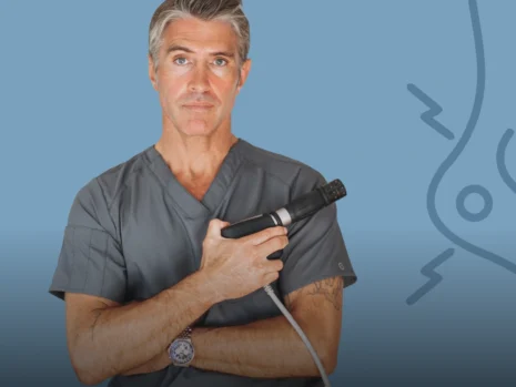
SHOCKWAVE THERAPY AND ACHILLES TENDONITIS
Will shockwave therapy (SWT) assist with Achilles tendonitis?
SWT is an extremely reliable treatment of choice for patients with A.T and other causes of heel pain. The treatment will reduce the symptoms but more importantly will increase the rate of recovery. As the sound waves stimulate the Achilles tendon, they cause micro-trauma and this increases blood flow to the affected area. Research has shown that there is an increase in small blood capillaries in the soft tissue surrounding the area being treated.
Ultrasound imaging has been carried out before and after shock wave therapy treatments and this has proven that treatment stimulates the formation of new blood vessels. It is also suggested that in patients with A.T the shockwave therapy breaks down scar tissue and stimulates new healthy tissue such as collagen.
During the treatment, the sound waves create physiological change to the nerve endings and this has a positive effect by reducing the pain. While pain relief is a welcome side effect in patients with A.T, it is not the primary reason for carrying out the treatment. Primarily, treatment is applied to the Achilles tendon in order to promote healing. Patients receiving a course of shock wave therapy will normally have one treatment per week for approximately 4 to 6 weeks.
The success rate of SWT in the treatment of A.T and other conditions such as plantar fasciitis is high. In combination with some other treatments and guidance, there is usually a complete recovery and a speedy drop-off in symptoms.
On occasions, the course of shockwave therapy will finish, and the patient may still feel some symptoms of A.T. However, this does not mean that the treatment has failed. The new blood vessels that have formed during treatment will continue to bring blood into the affected Achilles tendon area to promote healing. Within a number of weeks, the patient will make a full recovery from their Achilles tendon injury and will report that the symptoms have subsided completely.
Patients with A.T and other heel pain conditions, especially those with plantar fasciitis heel pain, are able to tolerate treatment from the shock wave therapy machine without too much discomfort. While the machine does apply pressure to the affected area, the vibrations from the machine are normally quite gentle during the initial phase of the session.
As the treatment progresses the vibrations from the machine have an analgesic effect on the nerve endings in the affected area and this creates a welcomed numbing sensation. This allows the practitioner to increase the pressure slightly so that the treatment is more effective. The shock wave therapy treatment continues in this fashion for approximately 3 to 4 minutes.
Following treatment, patients with A.T report instant relief and a significant improvement in symptoms as they stand and walk inside the consultation room.
ACHILLES TENDON TREATMENT
In order to explain some of the treatment options for A.T /tendinosis please refer to the following case study.
In September 2018 a healthy female individual, 38 years of age, arrived at the Sydney Heel Pain Clinic complaining of pain in the Achilles tendon area of her right ankle. She had been in pain for approximately 12 weeks and reported symptoms consistent with A.T. This lady had been to see her local physiotherapist and had been under her care for 6 weeks.
During the six-week period, the physiotherapist provided stretching and strengthening exercises and carried out deep tissue massage around the calf muscle area in an attempt to relieve stress on the Achilles tendon.
The patient reported a mild improvement in symptoms during the treatment and for the two days that followed but experienced a significant increase in her symptoms during and after the exercises that she was instructed to carry out.
This lady reports to the sports podiatrist that because her A.T was not improving, she decided to take a short course of medication to reduce the pain and inflammation. She also decided to stop running and refrained from any physical activity that would load the Achilles tendon. Ordinarily, this lady would carry out approximately 4 sessions of exercise each week and this would involve middle distance running of approximately 10 to 15 km.
As is common in patients with A.T, this patient advised the practitioner that she was able to carry out her physical activity without feeling too much pain. The first five minutes of exercise were difficult whereby she would feel stiffness and sharp pains through the Achilles tendon, but these symptoms would subside as she continued to run. Within approximately an hour of completing her run the typical symptoms of the A.T would return.
She would feel a sharp stabbing sensation through the shaft of the Achilles tendon and would be aware of heat and mild throbbing around the back of the ankle. The following morning when rising from bed, the classic A.T symptoms such as stiffness and stabbing pains would cause her to walk with a significant limp for at least 5 to 10 minutes. Throughout the day pain levels were manageable.
In order to relieve some of the symptoms, she would carry out some gentle massage around the Achilles tendon. She would also apply ice packs to the area whenever possible. A.T does involve some inflammatory change within the Achilles tendon, hence the patient found short-term relief from the application of cold packs.
PHYSICAL ASSESSMENT FOR ACHILLES TENDONITIS.
The sports podiatrist carried out a physical assessment with the patient lying prone on the treatment table, in order to measure the severity of the A.T. Visibly, the right Achilles tendon appeared to be larger and slightly thickened in comparison to the left. There was slight redness and mild heat. Lateral pressure was applied to the Achilles tendon using finger and thumb and this induced a significant jump response from the patient, confirming A.T.
Further down the Achilles tendon at the insertion onto the back of the heel bone pressure was applied without a response from the patient. This would exclude Achilles tendinosis in the most common part of the foot. The patient was asked to perform a single-leg heel raise which she managed albeit with mild pain. Tendon rupture was excluded from the diagnosis.
BIOMECHANICAL ASSESSMENT
The sports podiatrist carried out a biomechanical assessment with the patient walking and running on the treadmill, in order to exclude biomechanical anomalies as the cause of her A.T. Using sports podiatry techniques, her gait cycle was captured using digital software on an iPad. Anatomical landmarks were highlighted on the leg, Achilles tendon, and heel area using black texter.
The footage was replayed in slow motion and the following findings were noted:
- Early heel lift – right leg only.
- Significant pronation left foot, mild pronation right foot.
- Trendelenburg sign – right-sided hip drop/foot stomp. Indicating a right short leg.
- Mild genu valgum.
- Low cadence/increased stride length. Leading to a heavy heel strike.
ACHILLES TENDONITIS TREATMENT
The sports podiatrist explained to the patient that the likely cause of her A.T was an anatomical short right leg. It was explained that the short leg on the right side would lead to an early lifting of the right heel via the pulling of the calf muscle through the Achilles tendon. To this end, the patient was referred for a low radiation CT Scannogram to measure the length of her legs.
The result of the CT scan revealed an approximate difference of 12 mm (8 mm in the tibia, and 4 mm in the femur). The sports podiatrist explained to the patient the importance of using a heel left of approximately 6 to 8 mm inside the shoe of her right foot only. This would attempt to equalise her leg length which would contribute to an equal hip height and relieve stress on the lower back in addition to the knee. Importantly, the heel lift would reduce tension on the Achilles tendon which would reduce the repetitive stress in the affected area.
In order to treat the inflammation associated with this A.T, the patient was advised to continue with the use of ice packs on a daily basis. She was also advised to carry out specific calf muscle stretches at least three times a day. This would further reduce the stress on the Achilles tendon which would allow healing of the A.T.
The patient was using appropriate running shoes, but she was advised to avoid flat and flexible street shoes throughout the day. Footwear with a 10 or 20 mm raise at the heel would be more beneficial if the Achilles tendon was to heal quickly.
The patient was booked in for a three-week review in order to assess the improvement in the Achilles tendon. The use of shockwave therapy and acupuncture were discussed but ultimately were not necessary. At the 3-week follow-up appointment, the patient reported an improvement of approximately 70%. She had not returned to her usual running regime but reported to the sports podiatrist that the typical A.T symptoms that she felt after being seated or when rising from the bed were much more bearable. She was confident that her condition was improving and agreed not to proceed with further treatments. She was asked to return to the clinic for further assessment if her condition stopped improving or deteriorating. A follow-up appointment was made but the patient called the clinic to cancel her appointment as she was confident that she did not need further treatment.
She was advised to return to exercise slowly and to ensure at least two to three rest days in-between days when she ran, in order to ensure the A.T did not flare up. She was asked to carry out short runs of approximately 3 to 5 kilometres before increasing to her desired 10 to 15ks.
She was advised to maintain a strict calf stretching program at all times but in particular on the days of physical activity. She reported no negative side effects to the use of the left inside her right shoe.
Please note that the information in this case study should not be taken as general advice. If you think you have A.T or plantar fasciitis you should seek the help of a suitably qualified sports podiatrist or another medical practitioner.
Written by Karl Lockett
Contact Us To Find Out More About Achilles Tendonitis Treatments