Sydney Heel Pain Clinic

Heel Spurs

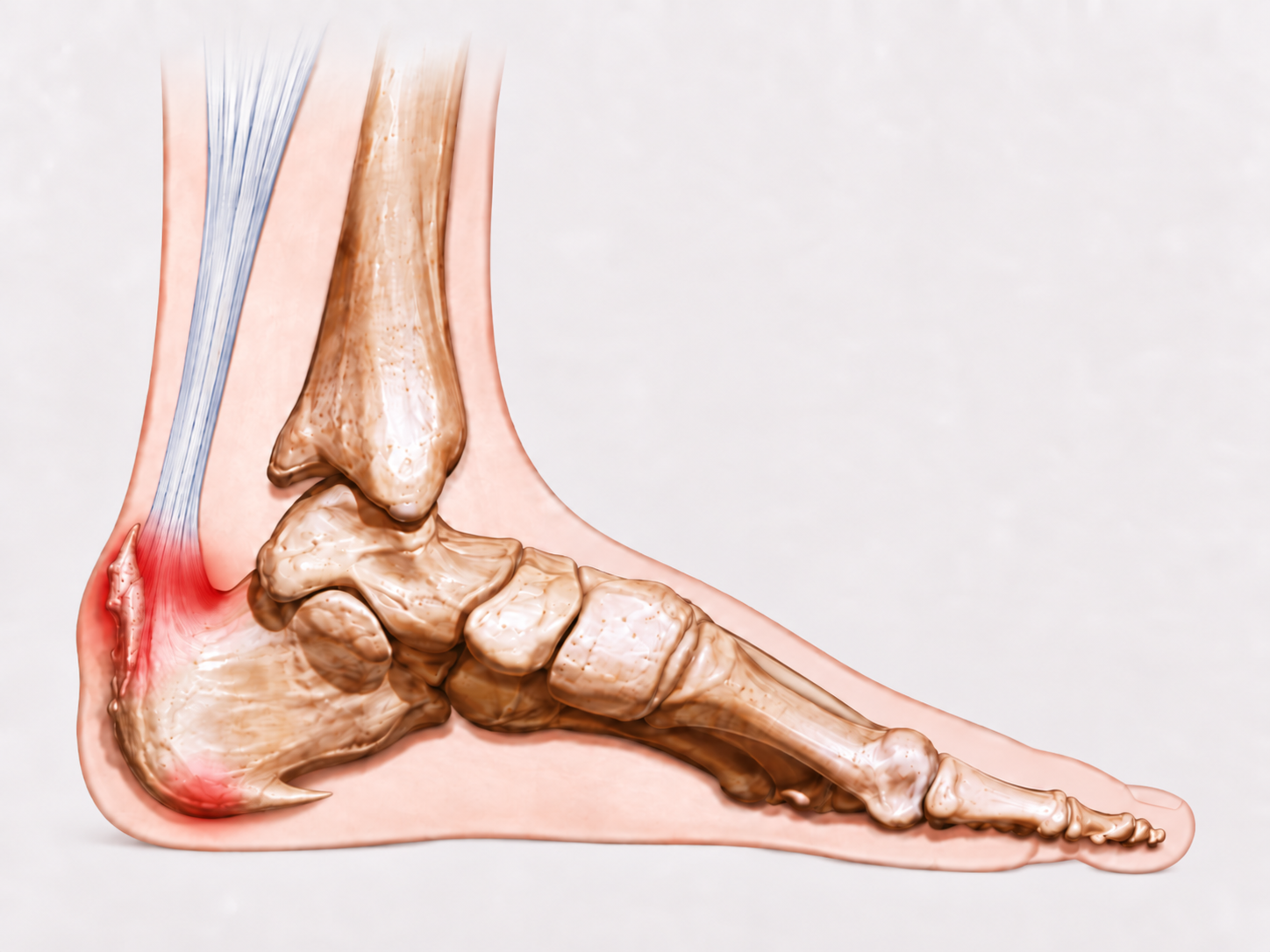
Heel spurs can develop underneath the heel or around the back of the calcaneus. Despite their appearance on X-ray, the spur itself is not usually the primary source of pain.

In most patients, symptoms arise from irritation or pathology within the soft tissues attached to, or positioned around, the heel bone. The clinical priority is therefore to identify the symptomatic tissue rather than assume that the bony spur is responsible.

Plantar and posterior calcaneal heel spurs with surrounding soft tissue irritation

Does This Sound Familiar?

Symptoms Associated With Heel Spurs

Symptoms differ according to whether the affected region is underneath the heel or around the posterior calcaneus.

Posterior Heel Spur

Pain at the Back of the Heel

  • Pain around the Achilles tendon insertion
  • Stiffness or discomfort following rest
  • Tenderness when pressure is applied to the back of the heel
  • Swelling or thickening around the Achilles insertion
  • Discomfort when the heel rests against a firm surface
  • Pain from footwear rubbing or compressing the back of the heel
Plantar Heel Spur

Pain Underneath the Heel

  • Pain concentrated beneath the heel bone
  • Marked discomfort with the first steps in the morning
  • Pain when standing following prolonged sitting
  • A deep bruised or stone-like sensation beneath the heel
  • Pain during prolonged standing or walking
  • Symptoms that can extend from the heel into the medial arch

The Important Distinction

The Heel Spur Is Usually Not the Pain Generator

A heel spur can appear substantial on medical imaging without being the structure responsible for the patient's symptoms.

Plantar Heel Spurs

Underneath the Heel

A plantar calcaneal spur can appear sharp and prominent on an X-ray. It is therefore understandable that patients frequently assume that the spur itself must be producing the pain.

In most cases, however, the symptomatic structure is the plantar fascia rather than the spur.

Plantar fasciitis develops around the attachment of the plantar fascia to the calcaneus, often in the same anatomical region in which the spur is visible.

Once the plantar fascia has recovered, symptoms can resolve completely even though the heel spur remains visible on X-ray.

Posterior Heel Spurs

At the Back of the Heel

Posterior calcaneal spurs develop close to the insertion of the Achilles tendon. Their presence does not automatically establish that the bone itself is responsible for the pain.

Symptoms in this region are frequently associated with insertional Achilles pathology, bursal irritation or compression of the surrounding soft tissues.

Clinical examination therefore needs to establish whether the symptoms originate from the Achilles insertion, a bursa, adjacent soft tissues or, less commonly, direct mechanical pressure from a substantial bony prominence.

The presence of a heel spur on an X-ray does not establish the source of pain. The symptomatic soft tissue must be identified clinically.

Associated Soft-Tissue Pathology

Conditions Commonly Responsible for the Pain

Heel spurs frequently coexist with pathology within structures attached to, or positioned around, the calcaneus.

Plantar Heel

Plantar Fasciitis

The most common source of pain associated with a plantar heel spur is pathology within the plantar fascia at its attachment beneath the calcaneus.

Plantar fasciitis →
Posterior Heel

Insertional Achilles Tendinopathy

Pathological change within the Achilles tendon close to its calcaneal insertion can produce focal pain, stiffness, thickening and compression sensitivity.

Insertional Achilles tendinopathy →
Posterior Heel

Retrocalcaneal Bursitis

Irritation of the bursa positioned between the Achilles tendon and calcaneus can produce deep posterior heel pain, particularly where local compression is increased.

Retrocalcaneal bursitis →
Plantar and posterior heel spurs can therefore coexist with different soft-tissue conditions. The clinical question is not simply whether a spur is present, but which tissue is symptomatic and what is maintaining irritation through that tissue.

Sydney Heel Pain Clinic

A Clinic Built Around Heel Pain

Heel pain cannot be diagnosed from the presence of a spur alone. Examination must identify the symptomatic tissue and establish the mechanical factors maintaining stress and irritation through that structure.

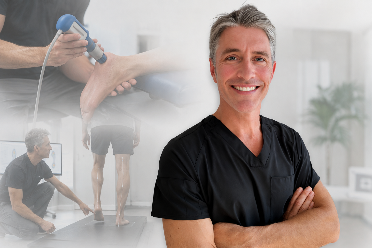
Sydney Heel Pain Clinic combines investigative clinical assessment, biomechanical investigation, gait assessment, footwear analysis and medical imaging where clinically indicated.

Heel pain assessment and treatment at Sydney Heel Pain Clinic

Establish Which Structure Is Responsible for the Pain

A heel spur is one clinical finding. Assessment determines whether symptoms arise from the plantar fascia, Achilles insertion, bursal structures, surrounding soft tissues or direct mechanical compression from the calcaneus.

Medical Imaging

When Is Imaging Required?

Medical imaging can be useful when symptoms persist, the diagnosis remains uncertain or the relationship between bony anatomy and the surrounding soft tissues requires further investigation.

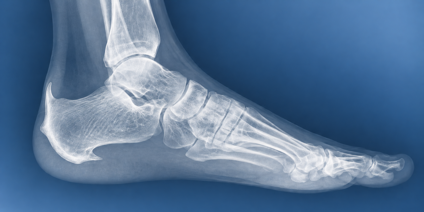
Lateral X-ray demonstrating plantar and posterior calcaneal heel spurs

X-ray

X-ray imaging demonstrates calcaneal bone formation and allows the position, orientation and size of plantar and posterior heel spurs to be assessed.

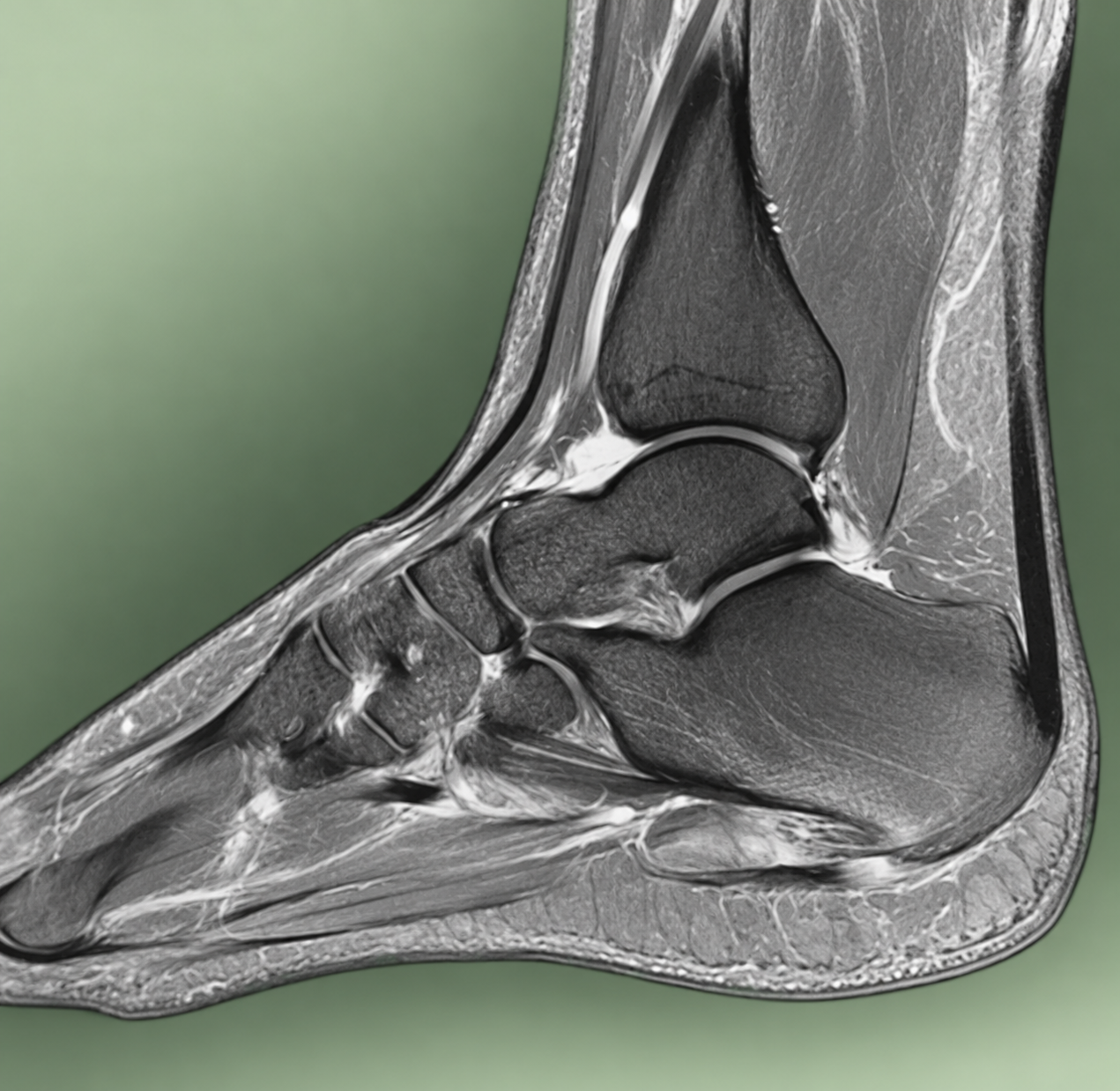
MRI imaging of the plantar fascia, heel bone and surrounding soft tissues

Ultrasound or MRI

Ultrasound or MRI can confirm plantar fascia pathology consistent with plantar fasciitis and assess tissue thickening, degeneration, inflammation or structural change where further investigation is clinically indicated.

Treatment

Treat the Painful Soft Tissue First

In most patients, the heel spur itself does not require treatment. Management is directed towards the symptomatic soft tissue and the mechanical forces maintaining stress through it.

The first priority is to identify the mechanical drivers maintaining persistent stress and irritation through the plantar fascia, Achilles tendon or surrounding soft tissues.

Treatment then focuses on reducing tissue irritation and inflammation, managing load and assessing tissue tolerance while removing or modifying the mechanical forces that are preventing recovery.

Depending on the clinical findings, this may involve modification of activity or loading, footwear recommendations, shockwave therapy to stimulate healing in selected chronic presentations, or custom orthoses where foot mechanics are contributing to persistent biomechanical stress.

In most cases, symptoms resolve once the painful soft tissue has recovered, even though the heel spur itself remains.

A Heel Spur on X-ray Does Not Automatically Require Treatment

The clinical priority is to establish whether the spur is an incidental finding or whether symptoms arise from pathology within the surrounding soft tissues or direct mechanical compression from a substantial bony prominence.

Surgical Intervention

When Can a Heel Spur Require Surgery?

Surgical intervention is considerably more relevant to selected posterior calcaneal abnormalities than to routine plantar heel spurs. Plantar heel spurs themselves rarely require surgical removal.

At the back of the heel, however, a substantial bony prominence can become clinically significant when its size and position create persistent compression against footwear or interfere mechanically with the Achilles tendon insertion.

Large Posterior Calcaneal Prominences

A prominent posterosuperior calcaneus, including a Haglund-type deformity, can create significant mechanical compression between the posterior heel, Achilles region and footwear.

Substantial spur formation extending into or around the Achilles insertion can also alter the contour and available space around the tendon, affecting normal tendon movement and flexibility.

Where there is persistent insertional Achilles pathology, substantial bony prominence and symptoms that remain despite appropriate conservative management, surgical assessment may be required.

Surgery is therefore considered after the symptomatic soft tissues, relevant mechanical drivers and conservative treatment options have been appropriately investigated and addressed.

Frequently Asked Questions

Heel Spur FAQs

Does the heel spur cause my pain?

Usually not. Underneath the heel, symptoms are commonly caused by plantar fasciitis affecting the plantar fascia around its attachment to the calcaneus.

At the back of the heel, symptoms frequently arise from the Achilles insertion, a bursa or adjacent soft tissues. Patients can become completely pain-free following treatment even though the heel spur remains.

How do I know if I have a heel spur?

Heel spurs are clearly demonstrated on X-ray. However, the presence of a spur does not establish that it is responsible for the symptoms. Clinical examination is required to identify the painful structure and determine whether the spur has any clinical relevance.

Can a heel spur disappear after treatment?

No. Once established, the bony spur generally remains. Successful treatment does not depend on removing the spur. Treatment is directed towards resolving the pathology and mechanical irritation affecting the symptomatic soft tissue.

Can heel spurs be surgically removed?

Yes, although surgical removal of a routine plantar heel spur is uncommon. Surgery is more relevant to selected posterior heel abnormalities where substantial bone formation produces persistent mechanical compression, significant Achilles insertional involvement or symptoms that remain despite appropriate conservative treatment.

Is a heel spur the same as plantar fasciitis?

No. A heel spur is bone formation arising from the calcaneus. Plantar fasciitis involves pathology of the plantar fascia at its attachment beneath the heel. The two can coexist but they are not the same condition.

Written by Karl Lockett, Sports Podiatrist

Investigative Assessment for Plantar and Posterior Heel Pain

Clinical assessment identifies the symptomatic tissue, investigates the mechanical factors maintaining irritation and determines whether the heel spur itself has any clinical significance.

Scroll to Top